The Neural
Control of Visually Guided Eye Movements
C. Cortical Mechanisms of Visually Guided Saccadic Eye Movements
 The
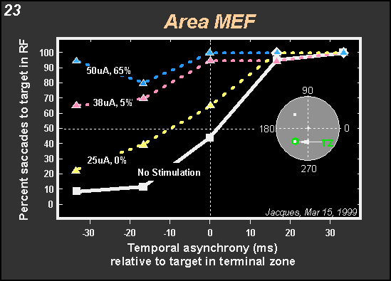
data shown in Figure 23 were obtained
while stimulating the medial eye fields (MEF). Stimulation consistently
produced facilitation as long as one of the targets was placed into the
terminal zone of the stimulated neurons. Since in MEF we have an orbital
code, the terminal zone is fixed in space as long as the head is stationary.
It is therefore possible to move the entire display so as to place the
fixation spot into the terminal zone instead of a target. When that is
done, stimulation causes a delay in saccade initiation by increasing fixation
time in a fashion similar to that found when those regions of LIP are
stimulated that contain fixation cells. Thus areas LIP and MEF appear
to play a role in decisions about when to initiate a saccadic
eye movement. The
data shown in Figure 23 were obtained
while stimulating the medial eye fields (MEF). Stimulation consistently
produced facilitation as long as one of the targets was placed into the
terminal zone of the stimulated neurons. Since in MEF we have an orbital
code, the terminal zone is fixed in space as long as the head is stationary.
It is therefore possible to move the entire display so as to place the
fixation spot into the terminal zone instead of a target. When that is
done, stimulation causes a delay in saccade initiation by increasing fixation
time in a fashion similar to that found when those regions of LIP are
stimulated that contain fixation cells. Thus areas LIP and MEF appear
to play a role in decisions about when to initiate a saccadic
eye movement.
|